Journal of Case Reports and Reviews in Medicine (ISSN: 3069-0749)
Case Report Volume: 2 & Issue: 3
















 <
<



Case Report Volume: 2 & Issue: 3
Background: Marginal ulcers following pancreaticoduodenectomy with Billroth II reconstruction are uncommon but may cause substantial morbidity when refractory to optimized medical therapy. Surgical revision is challenging because of altered anatomy, dense adhesions, and previous open surgery. Published evidence specifically addressing robotic revisional reconstruction for this late complication remains sparse.
Case Presentation: A 43-year-old man presented with persistent epigastric pain that was refractory to prolonged high-dose proton pump inhibitor therapy four years after pancreaticoduodenectomy with Billroth II reconstruction for a pancreatic head cyst. Upper gastrointestinal endoscopy demonstrated a chronic marginal ulcer at the gastrojejunostomy. Cross-sectional imaging excluded recurrence, anastomotic stricture, and other intra-abdominal pathology. Following multidisciplinary evaluation and discussion of open, laparoscopic, and robotic options, robotic-assisted revision was selected.
Results: The patient underwent robotic-assisted partial gastrectomy with conversion to Roux-en-Y reconstruction. Extensive adhesiolysis was required. Intraoperative indocyanine green (ICG) fluorescence angiography was used as an adjunct to confirm visually satisfactory perfusion of the reconstructed segments; it did not prompt a change in the planned reconstruction. Operative time was 120 minutes, with estimated blood loss <50 mL. Postoperatively, gastrojejunal anastomotic bleeding occurred and was successfully managed with endoscopic hemostasis (Clavien-Dindo grade IIIa). The patient was discharged on postoperative day 7, tolerating an oral diet.
Conclusion: Robotic-assisted conversion from Billroth II to Roux-en-Y reconstruction may be a feasible option for carefully selected patients with refractory marginal ulcer after pancreaticoduodenectomy when performed by an experienced minimally invasive team. In this case, ICG fluorescence was used as an adjunctive perfusion assessment tool, although a single case cannot establish a clinical benefit. Larger series with longer follow-up and comparative cost-eff ectiveness data are required to validate these observations.
keywords: Marginal ulcer; Pancreaticoduodenectomy; Billroth II reconstruction; Roux-en-Y reconstruction; Robotic surgery; Indocyanine green fluorescence angiography
Pancreaticoduodenectomy remains a standard resection for selected benign and malignant lesions of the pancreatic head and periampullary region. Contemporary open, laparoscopic, and robotic approaches have been described, with technique selection influenced by patient factors, disease characteristics, institutional volume, and surgical expertise.1,8 Billroth II gastrojejunostomy remains one reconstructive option but may predispose to bile reflux and marginal ulceration through chronic exposure of the gastric mucosa to biliary and pancreatic secretions.2,3
Although most marginal ulcers respond to medical therapy, a subset of patients develops persistent or recurrent symptoms that substantially impair quality of life and may require surgical revision.4 Proposed mechanisms include chronic bile reflux, altered mucosal defense, acid exposure, and, in selected patients, increased acid secretion related to altered postgastrectomy physiology. Conversion to Roux-en-Y reconstruction can divert bile away from the gastric remnant and has been associated with favorable outcomes in refractory disease.5
Reoperative surgery after pancreaticoduodenectomy is particularly challenging because of adhesions, distorted anatomy, and the proximity of the pancreaticojejunostomy and hepaticojejunostomy. Robotic assistance may provide three-dimensional visualization, wristed instrumentation, and stable dissection in confined spaces; however, these potential advantages must be balanced against cost, platform availability, and a substantial learning curve.8–10 Reports specifically describing robotic conversion of Billroth II to Roux en-Y for refractory marginal ulcer after pancreaticoduodenectomy are exceptionally limited. This case therefore contributes a detailed example of robotic reoperative reconstruction combined with fluorescence perfusion assessment and documents the management of a clinically significant postoperative bleeding complication.
A 43-year-old man presented with persistent moderate-intensity epigastric pain that was only partially responsive to prolonged high-dose proton pump inhibitor therapy and prokinetic agents. Symptoms had progressively worsened over several months and substantially affected quality of life.
The abdominal pain was predominantly postprandial, with worsening after meals, and was not associated with significant nocturnal symptoms. The patient reported no nausea or vomiting and had no clinical evidence of gastrointestinal bleeding, including hematemesis, melena, or hematochezia. Empirical treatment with proton pump inhibitors and antacids provided no significant symptomatic improvement. Prokinetic therapy was also ineffective.
Four years earlier, the patient had undergone pancreaticoduodenectomy for a pancreatic head cyst. The operation was initially attempted robotically but required conversion to open surgery because of intraoperative bleeding. Reconstruction was performed using a Billroth II gastrojejunostomy.
Upper gastrointestinal endoscopy revealed a chronic marginal ulcer at the gastrojejunostomy. Preoperative cross-sectional imaging three months before the proposed surgical intervention demonstrated the expected postoperative anatomy and excluded tumor recurrence, anastomotic stricture, a new mass, or other intra-abdominal pathology that could explain the symptoms. Given persistent symptoms despite optimized medical therapy, surgical revision was considered.
Preoperative laboratory evaluation showed a hemoglobin level of 13.0 g/dL and a serum albumin level of 3.7 g/dL. The patient’s body mass index was 21 kg/m². Helicobacter pylori testing was negative.
Case Selection and Justification
experience in pancreatic and minimally invasive surgery. Open, laparoscopic, and robotic revision were considered. Despite the previous conversion to open surgery and the anticipated adhesive burden, the robotic approach was favored because the pathology was localized to the gastrojejunal reconstruction, cross-sectional imaging showed no recurrent disease requiring oncologic resection, and the operating team had advanced experience in robotic foregut and revisional surgery. The patient was counseled regarding the possibility of difficult adhesiolysis and conversion to open surgery and expressed a preference for a minimally invasive approach after discussion of the potential risks and benefits.
The case was discussed at a multidisciplinary meeting involving gastrointestinal surgeons with extensive experience in robotic surgery, a general physician, and an experienced endoscopist. The patient’s previous surgical history, the anticipated complexity of the reoperative dissection, and the potential benefits and limitations of the available surgical approaches were carefully considered. After discussion of the therapeutic options with the patient, a robotic approach was selected. The decision was supported by the surgical team’s experience in advanced minimally invasive and robotic surgery. In this complex reoperative setting, the robotic platform was considered particularly advantageous because of its enhanced three-dimensional visualization, instrument articulation, and precision during adhesiolysis, dissection, and reconstruction.
A robotic-assisted approach was selected to address the anticipated technical complexity of reoperative upper abdominal surgery. After induction of general anesthesia, pneumoperitoneum was established and systematic exploration was performed using the da Vinci Xi (Intuitive Surgical, Sunnyvale, CA, USA) system.
Port configuration consisted of four 8-mm robotic trocars and one 12-mm assistant trocar. The robotic ports were distributed across the upper abdomen to provide adequate exposure and triangulation of the operative field, while the 12-mm assistant port was positioned to facilitate suction, retraction, and introduction of additional instruments as required. The robotic system was docked for an upper abdominal approach.
Extensive adhesions were encountered in the upper abdomen, involving the gastric remnant, transverse colon/mesocolon, jejunal limbs, and anterior abdominal wall around the previous operative field. These adhesions obscured the prior gastrojejunostomy and required meticulous adhesiolysis to restore the anatomy while avoiding injury to the existing pancreatic and biliary reconstructions. The previous Billroth II gastrojejunostomy was then identified.
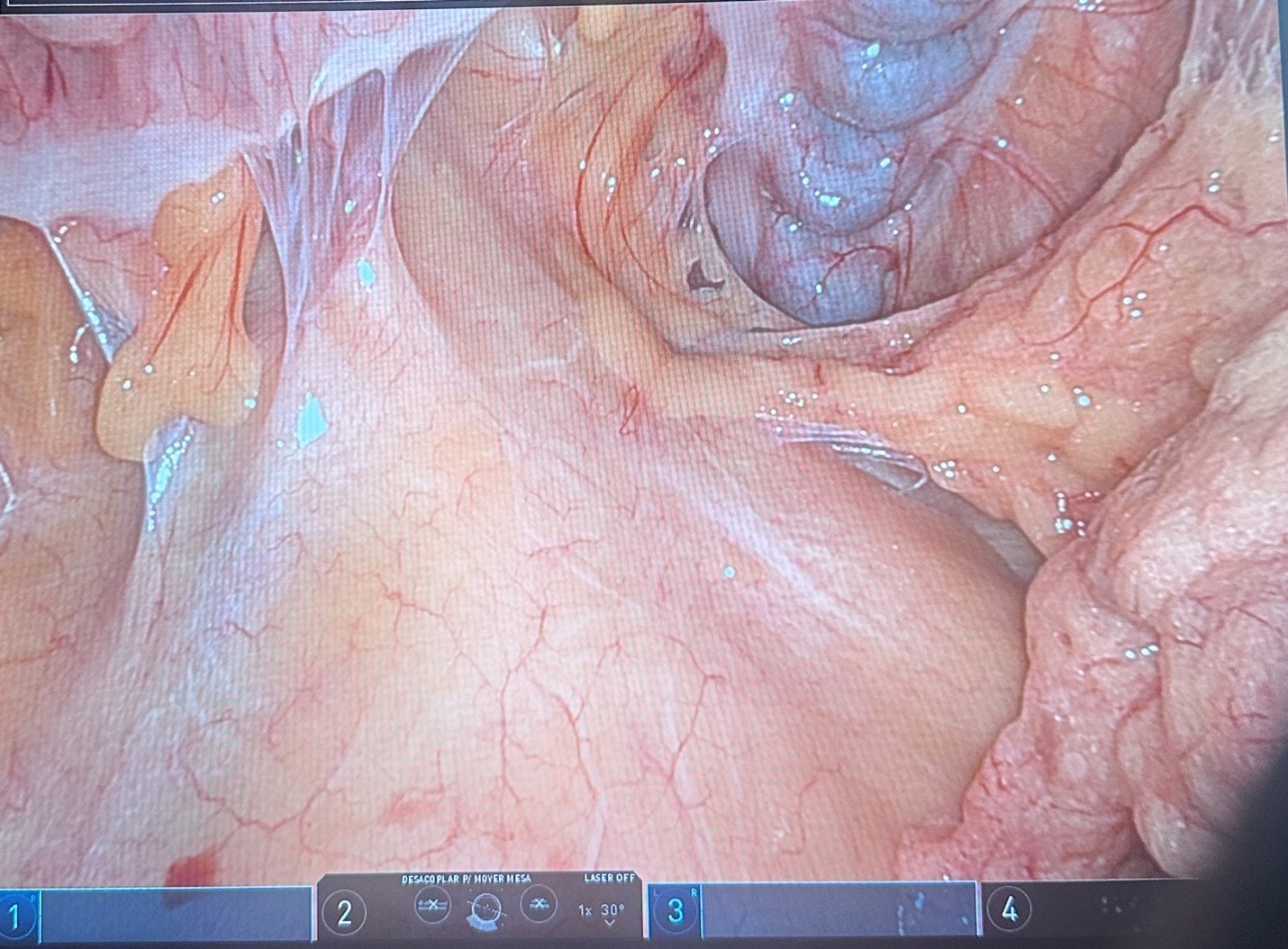
Figure 1. Extensive upper-abdominal adhesions involving the gastric remnant, jejunal limbs, transverse colon/mesocolon, and the previous operative field, requiring careful robotic adhesiolysis to expose the prior Billroth II gastrojejunostomy.
The operative strategy consisted of partial gastrectomy with conversion to Roux-en-Y reconstruction. The gastric antrum was transected using 60-mm linear stapler cartridges. The jejunum was divided proximal and distal to the previous gastrojejunostomy, thereby dismantling the Billroth II configuration.
A new gastrojejunostomy was constructed using the alimentary limb, followed by a side-to-side jejunojejunostomy 100 cm distal to the gastrojejunostomy to complete the Roux-en-Y reconstruction. All mesenteric defects were carefully closed using barbed sutures to reduce the risk of internal herniation.
The pancreaticojejunostomy and hepaticojejunostomy were carefully inspected intraoperatively and found to be intact, without evidence of anastomotic disruption or other abnormalities requiring surgical intervention. Neither anastomosis required revision.
Intraoperative indocyanine green (ICG) fluorescence angiography was used to assess perfusion of the reconstructed segments (Figure 2). Fluorescence demonstrated prompt and homogeneous perfusion of the new gastrojejunostomy and adjacent bowel. In this case, the findings supported the planned reconstruction but did not alter the transection level or require revision of an anastomosis.6,7
ICG was administered intravenously as a 5-mg bolus, followed by a saline flush. Near-infrared fluorescence imaging was performed using the Firefly® fluorescence imaging system integrated into the da Vinci Xi platform to assess perfusion of the newly constructed gastrojejunostomy and reconstructed bowel segments.
Total operative time was 120 minutes, and estimated blood loss was less than 50 mL.
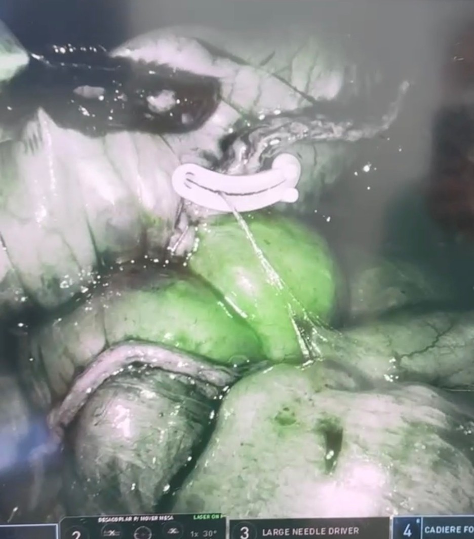
Figure 2. ICG fluorescence assessment of the newly constructed gastrojejunostomy, demonstrating prompt and homogeneous perfusion of the gastric and jejunal sides without a visible perfusion defect. The fluorescence findings did not prompt revision of the anastomosis.
The postoperative course was complicated by bleeding from the gastrojejunal anastomosis. Hemoglobin decreased from 12.4 g/dL to 11.1 g/dL. Urgent upper gastrointestinal endoscopy revealed an adherent clot and an exposed vessel at the gastrojejunostomy, with intermittent active bleeding. Complete hemostasis was successfully achieved by endoscopic clip placement. No blood transfusion was required. Anastomotic leakage was excluded. The event was classified as Clavien–Dindo grade IIIa.
After endoscopic hemostasis, recovery was uneventful. The patient progressively resumed oral intake and was discharged on postoperative day 7 in good clinical condition.
At follow-up, the patient demonstrated an excellent clinical outcome, with complete resolution of abdominal pain and no recurrence of symptoms. His nutritional status remained satisfactory, with a body mass index of 22 kg/m². Proton pump inhibitor therapy was continued for 60 days postoperatively and subsequently discontinued. Follow-up upper gastrointestinal endoscopy performed after completion of PPI therapy showed no abnormalities, with complete healing of the previously documented anastomotic ulcer and no evidence of recurrent ulceration.
Postoperative recovery was satisfactory. The patient passed flatus on postoperative day (POD) 3, indicating return of bowel function. No enteral or parenteral nutritional support was required during the postoperative period. The surgical drain showed a low-volume serohematic output, approximately 15 mL, and was removed on POD 6. The patient was discharged on proton pump inhibitor therapy at a dose of 80 mg/day for the first 30 days, followed by 40 mg/day for an additional 30 days. Discharge medications also included non-opioid analgesics as needed and simethicone for three days.
Informed Consent
Written informed consent was obtained from the patient for the publication of this case report and all accompanying clinical and surgical images. The consent form was signed by the patient, explicitly authorizing the use and publication of the case-related information and images.
Marginal ulceration following Billroth II reconstruction is multifactorial and is commonly associated with chronic bile reflux, acid exposure, altered gastric physiology, and impaired mucosal defense.2,3 Changes in postgastrectomy physiology may also influence gastrin and acid secretion in selected patients. After pancreaticoduodenectomy, this complication may present years after the index operation and can be difficult to manage because of altered anatomy and dense adhesions.4
Conversion to Roux-en-Y reconstruction offers a physiological rationale by diverting bile away from the gastric remnant and has shown favorable outcomes in the management of refractory marginal ulceration.5 The present case is notable because the revision was performed robotically after a previous operation that had itself required conversion to open surgery. The literature on robotic reoperative reconstruction specifically for marginal ulcer after pancreaticoduodenectomy is sparse; therefore, this report should be interpreted primarily as a technical and feasibility observation rather than evidence of superiority.
In the present case, robotic assistance facilitated meticulous adhesiolysis and reconstruction in a scarred upper abdomen. Three-dimensional visualization and articulated instruments were particularly useful around the previous gastrojejunostomy and adjacent reconstructed anatomy. Nevertheless, a laparoscopic approach could also have been feasible in experienced hands. The robotic platform was favored because of the anticipated need for precise adhesiolysis and intracorporeal reconstruction and because of the operating team’s experience with robotic revisional surgery. These advantages must be balanced against higher acquisition and procedural costs, limited platform availability, and the learning curve associated with complex robotic pancreatic surgery.8–10 Comparative evidence does not establish universal superiority of robotic over laparoscopic surgery, and approach selection should remain individualized.9
ICG fluorescence angiography provided real-time visual confi rmation of perfusion at the newly constructed anastomosis.6,7 Importantly, fluorescence did not alter the planned transection level or prompt anastomotic revision in this case. Accordingly, no causal conclusion can be drawn regarding its effect on postoperative outcomes, and the subsequent anastomotic bleeding further illustrates that satisfactory fluorescence perfusion does not exclude non-ischemic complications. In a single case, ICG should therefore be regarded as an adjunctive assessment tool rather than a validated means of improving outcomes.
Strengths
Strengths of this report include the detailed description of a technically challenging revisional reconstruction, use of ICG fluorescence as an adjunctive intraoperative assessment tool, and documentation of successful endoscopic management of a clinically significant postoperative complication.
Limitations
This report is limited by its single-case design, which precludes generalization or comparative conclusions. Outcomes may be influenced by surgeon and institutional expertise, introducing selection and experience bias. In addition, no cost-eff ectiveness analysis was performed, and the clinical benefit of ICG fluorescence cannot be established from this observation.
Robotic-assisted conversion from Billroth II to Roux-en-Y reconstruction may be a feasible option for selected patients with refractory marginal ulcer after pancreaticoduodenectomy when performed by an experienced team. The robotic platform can facilitate dissection and reconstruction in a challenging reoperative field, while ICG fluorescence may provide adjunctive perfusion information. However, this single case cannot demonstrate superiority over laparoscopic or open revision or establish a clinical benefit from fluorescence imaging. Larger studies with longer follow-up and cost-effectiveness evaluation are needed to validate these observations.
