Journal of Case Reports and Reviews in Medicine (ISSN: 3069-0749)
Review Article Volume: 2 & Issue: 3




















Review Article Volume: 2 & Issue: 3
Ocular tuberculosis is a rare form of extrapulmonary tuberculosis that can affect virtually any ocular tissue. We report the case of a 15-yearold adolescent from a tuberculosis-endemic region who presented with unilateral redness of the left eye and blurred vision, without pain, photophobia, or systemic symptoms, and with no relevant medical history. The condition failed to respond to topical corticosteroid and non-steroidal anti-inflammatory treatment, prompting further investigation. A comprehensive infectious and autoimmune work-up was negative, while a tuberculin skin test (7 mm induration) and a QuantiFERON-TB Gold assay were both positive, despite normal chest radiography, negative sputum smear microscopy, and a negative GeneXpert MTB/RIF assay. A diagnosis of tuberculous scleritis was made, and the patient was treated with a standard four-drug antituberculous regimen combined with oral corticosteroids. Ocular redness resolved completely and vision improved within three weeks, with no recurrence at follow-up. This case illustrates that ocular tuberculosis should be considered in the differential diagnosis of persistent, treatment-resistant ocular inflammation in tuberculosis-endemic areas, even in the absence of pulmonary or systemic disease.
keywords: Ocular tuberculosis; tubercular scleritis; extrapulmonary tuberculosis; QuantiFERON-TB Gold; adolescent; case report.
Tuberculosis (TB) remains a major global public health concern, particularly in endemic regions. According to the World Health Organization’s 2024 Global Tuberculosis Report, an estimated 10.8 million people developed active tuberculosis in 2023, and the disease remained the leading cause of death from a single infectious agent worldwide.1 While pulmonary disease accounts for the majority of cases, extrapulmonary tuberculosis represents approximately 15–20% of the global burden, and ocular involvement is among its rarest manifestations.1
A recent systematic review and meta-analysis estimated the pooled global prevalence of tubercular uveitis at approximately 4% of all uveitis cases, rising to 7% in tuberculosis high-burden countries and up to 11% in sub-Saharan Africa.2 In low-incidence regions such as Europe and North America, ocular tuberculosis remains rare, and the risk is increased among immunocompromised individuals and those with HIV co-infection.3
Diagnosing ocular tuberculosis is challenging because of the diversity of its clinical presentations, which overlap with numerous other infl ammatory and infectious ocular conditions, and because no single confi rmatory test exists.3 Ocular tuberculosis may present as anterior or posterior uveitis, choroiditis with or without granuloma formation, retinitis, scleritis, tuberculous conjunctivitis, or optic neuropathy, occurring alone or in combination, and requires prompt recognition to prevent irreversible visual complications.4 We report a case of ocular tuberculosis presenting as unilateral scleritis in a 15-year-old adolescent from a tuberculosis-endemic area, to illustrate the diagnostic reasoning required when systemic and pulmonary evaluations are unrevealing.
Patient information
A 15-year-old female adolescent, resident in a tuberculosisendemic region of Madagascar, presented to the ophthalmology department in April 2024 with unilateral redness of the left eye. She had been vaccinated against tuberculosis with BCG and had no relevant medical history, including no recurrent oral aphthosis. She denied constitutional symptoms suggestive of active tuberculosis, including asthenia, anorexia, weight loss, night sweats, or fever.
The patient was initially treated empirically with a topical corticosteroid combination (Chibro-cadron, 5 drops daily) for 24 hours, without improvement. Treatment was then changed to topical indomethacin, a non-steroidal anti-inflammatory drug, 1 drop three times daily. Despite this change, the ocular redness and blurred vision persisted, prompting referral for further evaluation.
Clinical findings
On examination, the left eye was diffusely red, with dilation of the episcleral vessels and no pain or tenderness on palpation (Figure 1). Visual acuity was reduced in the affected eye, without other identifiable anterior- or posterior-segment abnormalities. The right eye was normal. General physical examination showed no clinical signs of active systemic tuberculosis.
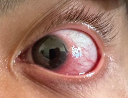
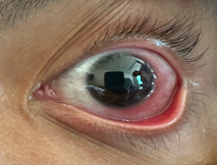
Figure 1. Diffuse anterior scleritis of the left eye at presentation, with localized redness and dilation of the episcleral vessels.
Timeline
| Time point | Event |
|---|---|
| April 2024 | Onset of unilateral left-eye redness and blurred vision; presentation to the ophthalmology department. |
| Days 0–1 | Empirical topical corticosteroid (Chibrocadron), 5 drops/day for 24 hours — no improvement. |
| Following days | Switch to topical indomethacin, 1 drop 3×/day — symptoms persist; referral for further work-up. |
| Work-up | Laboratory, serologic, and autoimmune panels unremarkable; TST positive (7 mm); QuantiFERON-TB Gold positive; chest X-ray normal; sputum AFB negative; GeneXpert MTB/RIF negative. |
| Diagnosis | Tuberculous scleritis (ocular tuberculosis) established on clinical, epidemiological, and immunological grounds. |
| Treatment start | Antituberculous therapy initiated (2-month intensive phase HRZE) with oral prednisone 0.5 mg/kg/day. |
| Week 3 | Complete resolution of ocular redness; progressive improvement of vision (Figure 2). |
| Months 2–6 | Continuation phase (isoniazid + rifampicin) completed; regular ophthalmologic follow-up; no recurrence. |
Laboratory investigations
Complete blood count, C-reactive protein, renal function, liver function, and serum electrolytes were within normal limits. Serologic testing for HIV, hepatitis B and C, syphilis, toxoplasmosis, and rubella was negative. Serum protein electrophoresis was unremarkable. Antinuclear antibodies, antidouble-stranded DNA antibodies, antineutrophil cytoplasmic antibodies (ANCA), and serum angiotensin-converting enzyme were negative, and HLA-B27 typing was negative, arguing against the principal autoimmune and HLA-B27-associated causes of scleritis considered in the differential diagnosis.
Tuberculosis-specific investigations
The tuberculin skin test produced a 7 mm induration, and the interferon-gamma release assay (QuantiFERON-TB Gold), which detects T-cell responses to Mycobacterium tuberculosis-specific antigens, was positive.5 Chest radiography was normal, sputum smear microscopy showed no acid-fast bacilli, and GeneXpert MTB/RIF testing of sputum was negative for M. tuberculosis DNA.
Diagnosis
Given the persistent, treatment-resistant ocular inflammation, the negative results of a broad infectious and autoimmune work-up, and the concordant positive tuberculin skin test and QuantiFERON-TB Gold assay, a diagnosis of tuberculous scleritis, within the spectrum of ocular tuberculosis, was made in this tuberculosis-endemic setting.6 The normal chest radiograph and negative sputum studies did not exclude the diagnosis, since extrapulmonary tuberculosis, including ocular disease, commonly occurs without demonstrable pulmonary involvement.
Therapeutic intervention
The patient was started on standard first-line antituberculous therapy according to the national tuberculosis control programme: an intensive phase of isoniazid, rifampicin, ethambutol, and pyrazinamide for two months, followed by a four-month continuation phase of isoniazid and rifampicin.12 Oral prednisone, 0.5 mg/kg/day, was added to control ocular inflammation.
Follow-up and outcome
By the third week of antituberculous treatment, the ocular redness had completely resolved and visual acuity was improving (Figure 2). No adverse effects or treatment-related complications were observed. The patient completed the six-month antituberculous regimen and remains in clinical remission, with regular ophthalmologic follow-up to monitor for recurrence or late complications.
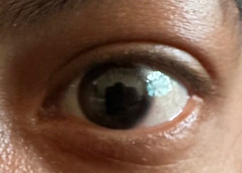
Figure 2. Complete resolution of ocular redness one month after initiation of antituberculous therapy.
Ocular tuberculosis is an uncommon but clinically important manifestation of Mycobacterium tuberculosis infection, particularly in endemic regions. Its diagnosis is often delayed because systemic symptoms are frequently absent and the clinical presentation overlaps with numerous other causes of ocular inflammation.
Tuberculosis remains one of the leading causes of death from a single infectious agent worldwide, with an estimated 10.8 million new cases in 2023.1 Extrapulmonary disease accounts for a substantial minority of cases, and ocular involvement is among its least common forms.4 Pooled estimates from a recent systematic review and meta-analysis indicate that tubercular uveitis affects approximately 4% of uveitis cases worldwide, rising to 7% in tuberculosis high-burden countries and up to 11% in sub-Saharan Africa, while remaining substantially rarer in lowincidence settings such as Europe and North America.2 Ocular tuberculosis can occur in immunocompetent individuals, as in the present case, although the risk is increased in those who are immunocompromised or HIV-co-infected.3 Adolescents and young adults are a recognized vulnerable group, often in the context of household or community exposure to tuberculosis.7
Clinically, ocular tuberculosis is protean, encompassing conjunctivitis, scleritis, keratitis, anterior or posterior uveitis, retinitis, and optic neuropathy, which may occur alone or in combination.6 Our patient presented with isolated unilateral scleritis, without pain, photophobia, or systemic symptoms, a pattern consistent with previously reported presentations of ocular tuberculosis in endemic areas.8
No single test can confirm ocular tuberculosis, and diagnosis relies on the convergence of clinical, epidemiological, and immunological evidence.9 The tuberculin skin test measures delayed-type hypersensitivity to tuberculin antigens,10 while interferon-gamma release assays such as QuantiFERON-TB Gold detect T-cell responses to M. tuberculosis-specific antigens and are increasingly used to support the diagnosis of latent or active infection.5 In this case, the concordance of a positive tuberculin skin test and a positive QuantiFERON-TB Gold assay, together with a compatible clinical picture and exclusion of alternative causes, supported the diagnosis despite a normal chest radiograph and negative sputum studies. The absence of pulmonary findings does not exclude ocular tuberculosis, which can occur without concurrent pulmonary disease.11
Treatment of ocular tuberculosis generally follows standard antituberculous regimens used for pulmonary disease: an intensive phase with isoniazid, rifampicin, ethambutol, and pyrazinamide, followed by a continuation phase with isoniazid and rifampicin.12 Systemic corticosteroids are frequently added to control the inflammatory response and reduce the risk of irreversible ocular damage,13 since inflammation associated with intraocular tuberculosis is thought to reflect both direct infection and a delayed hypersensitivity response to mycobacterial antigens.14 In our patient, the combination of antituberculous therapy and oral corticosteroids led to complete clinical resolution within three weeks, consistent with outcomes described in other reported series.15 Long-term ophthalmologic follow-up remains important, as patients with ocular tuberculosis can experience relapse or late complications such as cataract or secondary glaucoma.15
This report has limitations inherent to a single case, most notably the absence of histopathological or microbiological confirmation of M. tuberculosis within ocular tissue - a recognized diagnostic and technical challenge in ocular tuberculosis - and reliance on indirect immunological evidence alongside clinical response to antituberculous therapy. The favorable and rapid treatment response, together with exclusion of alternative infectious and autoimmune causes, nonetheless provides indirect support for the diagnosis.
This case adds to the limited pediatric and adolescent literature on ocular tuberculosis and reinforces that the diagnosis should be actively considered in tuberculosis-endemic regions, even when systemic and radiological evidence of tuberculosis is lacking.
Patient perspective
The patient and her family reported considerable concern about the persistent ocular redness and blurred vision prior to diagnosis, compounded by the lack of improvement with initial topical treatments. Following initiation of antituberculous therapy, they described rapid relief of symptoms and expressed satisfaction with the clarity of the explanations provided about the diagnosis and treatment plan.
This case of ocular tuberculosis illustrates the complexity of diagnosing extrapulmonary tuberculosis, particularly in the absence of systemic symptoms. It highlights the importance of considering tuberculosis in the differential diagnosis of persistent ocular inflammation, especially in regions where tuberculosis is endemic. Concordant positive results on the tuberculin skin test and QuantiFERON-TB Gold assay were key to establishing the diagnosis, despite a normal chest radiograph and negative sputum studies. The rapid clinical improvement following a tailored antituberculous regimen further supports the diagnosis.
Conflicts of interest
The authors declare that they have no conflicts of interest.
Authors’ contributions
AM Andrianiaina, RMF Randrianarisoa, P Raharitina and H Ramanandafy drafted the report. MI Rahatamalala contributed to the conception and critical revision of the report. HMD Vololontiana validated the report. All authors read and approved the final version of the manuscript.
Acknowledgments
The authors thank the internal medicine staff of Befelatanana and Anosiala hospitals.
Consent to publication
Written informed consent was obtained from the patient and her legal guardian to publish this report, in accordance with the journal’s patient consent policy. Only information necessary for scientific understanding was included, and the patient’s anonymity was preserved.
Data availability statement
All data generated or analyzed during this case are included in this article.
Funding
The authors declare that they received no funding from any specifi c organization for this work.
