Journal of Case Reports and Reviews in Medicine (ISSN: 3069-0749)
Review Article Volume: 2 & Issue: 3
















 <
<



Review Article Volume: 2 & Issue: 3
Squamous cell carcinoma (SCC) is a common cancer of the oral cavity. It mainly affects males more than females, with middle-aged to elderly people being the most susceptible. In this report, we present a case of an 88-year-old male with a mass on the posterior-lateral border of the tongue. After thorough clinical, histopathological, and imaging assessments, an SCC was confirmed. Because of the patient’s age, the oncologist has chosen immunotherapy over invasive options of surgery, radiotherapy, and chemotherapy.
keywords: cancer, oral, squamous cell carcinoma, tongue.
Squamous cell carcinoma (SCC) is a malignant epithelium-derived neoplasm that can develop all over the body but primarily in the skin and oral cavity;1 it accounts for up to 90% of all oral cancers and generally occurs in males more than females, with middle-aged to elderly people being the most susceptible.2-4 SCCs of the tongue, often found at the lateral border, represent 25 to 40% of these neoplasms. Along with SCCs of the floor of the mouth, they make up over 50% of intraoral carcinomas. The hard palate, gingiva, buccal mucosa, and soft palate are less likely to be aff ected.5,6 Oral SCC (OSCC) results in disfigurement and functional impairments, such as challenges with swallowing, speech, and taste, which significantly impact patients’ quality of life.7,8 Diagnosis of OSCCs needs thorough clinical, radiological, and histological evaluations; clinically, they are characterized by a lesion that appears red and white or solely red (erythroplakia), featuring a rough surface and distinct borders.9,10 At an early stage they are typically painless; however, they can lead to discomfort and may develop features such as ulceration, nodularity, and tissue attachment as they progress.2,11 Ulceration is a common symptom of OSCC, presenting with an irregular floor and raised hard exophytic margins, and is firm upon palpation.5,11,12 Radiologically, magnetic resonance imaging (MRI) and computed tomography (CT) are crucial for evaluating OSCC.5 Their main advantage lies in their ability to determine the extent of the lesion, the depth of invasion, the involvement of the neighboring muscles, and importantly, the characteristics of the lymph nodes, including their number, size, location, contour, and presence of necrosis.5,13 Histologically, the lesion begins as an epithelial dysplasia characterized by an altered proliferation of dysplastic squamous cells on the epithelium surface. This alteration leads to the degradation of the sub-epithelial basement membrane, resulting in local destruction and the potential for distant invasion through metastasis.14 Management of OSCC primarily involves surgical intervention, particularly radical neck dissection when lymph node involvement is present. Additionally, radiotherapy and chemotherapy are utilized as supplementary postoperative treatments for advanced stages of cancer.15 On the other hand, immunotherapy has shown significant therapeutic benefi ts for patients with metastatic or locally advanced tumors not eligible for surgery or radiotherapy to avoid the potential toxicity caused by the chemotherapies.16
The geriatric age group is often associated with comorbidities, which might make therapy implementation challenging. Elderly patients may have a decreased capacity for tissue healing, may struggle to comply with the treatment plan, and may not be able to tolerate extensive surgery, as well as aggressive radiotherapy and chemotherapy.17,18 For that, oncologists must evaluate older patients on an individualized, case-by-case basis before implementing a treatment.17
In this report, we discuss a case of SCC located on the posterior lateral border of the tongue of an 88-year-old male patient.
An 88-year-old male was referred to our specialized oral medicine clinic for evaluation of a mass on the left posterior-lateral border of the tongue. Medically, he suffered from a cerebral embolism ten years ago, which was treated at the time without any consequences or recurrence. Moreover, he was diagnosed three years back with a high grade papillary urothelial carcinoma, which invades the lamina propria and is classified as stage pT1 according to the American Joint Committee on Cancer (AJCC). Due to his age, he was treated solely with chemotherapy. Currently, he is on medication to manage uric acid levels. He used to smoke one pack of cigarettes for thirty years until quitting twenty years ago.
The patient presented a completely edentulous upper jaw and a bilateral posterior edentulous lower jaw upon intraoral examination. He reported experiencing a speech disturbance and having difficulty opening his mouth. The lesion was firm and exhibited an exophytic white surface with ill-defined indurated margins. It was fixed to the underlying tissues and extended inferiorly to the floor of the mouth and the alveolar ridge of the mandible. Antero-posteriorly, it spanned from the premolar region to the 2nd molar region (Figure 1). On neck palpation, there were enlarged, non-tender, and firm left cervical lymph nodes.
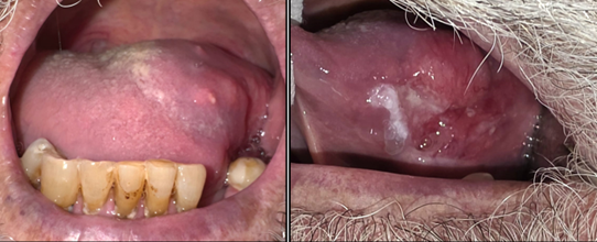
Figure 1. Proliferative mass featuring an exophytic white surface and ill-defined indurated margins, located in the left lateral border of the tongue, extending inferiorly to the floor of the mouth and the alveolar ridge of the mandible.
The panoramic radiograph demonstrates advanced partial edentulism with severe generalized alveolar ridge resorption, particularly in the maxilla. A limited number of mandibular teeth remain, predominantly in the anterior region, with advanced generalized periodontal bone loss. A mixed radiolucent/radiopaque image is present in the left posterior mandibular region partially extending in the ramus. The radiopacity is irregular in shape. There is no obvious radiographic evidence of mandibular cortical invasion in the region corresponding to the clinically described tongue lesion (Figure 2). Consequently, the definitive diagnosis cannot be established radiographically and requires histopathologic confirmation.
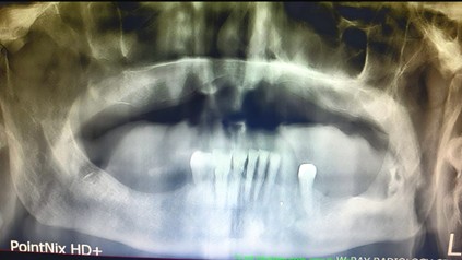
Figure 2. The mixed radiolucent/radiopaque image present in the left posterior mandibular region partially extending in the ramus.
A biopsy of the lesion was performed and sent for histopathological assessment. Microscopic examination showed a neoplastic keratinized proliferation consisting of moderately differentiated squamous cells arranged in trabeculae of variable size infiltrating the underlying lamina propria (Figure 3). The histological diagnosis of poorly differentiated SCC was made.
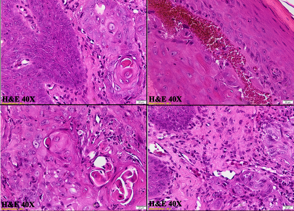
Figure 3. Microscopic features of the lesion.
A total-body positron emission tomography (PET)/CT scan was performed. A highly fluorodeoxyglucose (FDG)-avid left oral cavity mass (tongue) measuring 34x26 mm, strongly suspicious for a primary oral cavity malignancy. Additionally, FDG-avid left, mid-, and lower jugulo-carotid adenopathies, which were highly suspicious for nodal involvement, were detected.
The histopathological examination and PET scan have ruled out the presence of a metastatic tumor from the previous urothelial carcinoma.
The patient was referred to a head and neck oncologist, who, given his age, has proposed immunotherapy treatment.
SCC is the most common cancer of the oral cavity with male-to female ratio 2:1.5,13,15 Most of OSCC cases are related to tobacco and alcohol use. Furthermore, a study conducted by Hashibe et al. concluded that the combined effect between tobacco and alcohol use makes the head and neck cancer risk even higher.19 Additionally, several other factors have been proposed as risk factors, including ultraviolet light, human papillomavirus (HPV), candidiasis, radiation, genetic factors, malnourishment, diet, and chemical exposures to betel quit and areca nut.20,21
Before developing into the malignant stage, OSCCs usually begin as premalignant lesions, such as leukoplakia and erythroplakia, histologically characterized by degrees of dysplasia and clinically asymptomatic.5,15,21
OSCCs are frequently misdiagnosed or detected at later stages (III or IV), leading to delays in receiving timely and appropriate treatment. This results in a significant decline in patients’ quality of life and a noticeable reduction in their survival chances. Their five-year survival rates remain below 12%.14,22
Our patient is at risk for developing oral metastasis due to a history of urothelial carcinoma. Additionally, the initial lesion located on the posterior-lateral border of the tongue may have been ignored or underestimated in prior oral examinations.
The histopathological assessment of SCC establishes tumor grading by identifying abnormal cell differentiation. Poorly diff erentiated and metastasized SCCs are typically regarded as more aggressive and often have a worse prognosis.22 Additionally, it is usually accepted that better prognosis is related to early detected OSCCs.5,22 In our case, the lesion was moderately differentiated.
The management of OSCC in elderly patients requires collaboration among gerontologists, surgeons and oncologists due to its complexity. The severity of the illness and preoperative morbid conditions infl uence the therapy decision.
